
Scientist has formula for success:
Perseverance, perspective and a healthy sense of humor
Sometimes science is messy, and not for the faint of heart. It requires perseverance.
As a scientist currently immersed in groundbreaking work developing less-invasive diagnostic tests for cancer and Lyme disease, Joshua Nordberg ’00 knows what it means to persevere—even in the most trying of circumstances.
A sense of humor helps, which is something he learned from Professor of Biology Robert Morris. Like the time sea urchins started spawning in the lab aquarium, forcing a panicked Nordberg to call Morris at 1 a.m.
“Dr. Bob rushed back to the lab to clean up, laughing the whole time,” recalled Nordberg, a biology major. It was among their many funny, legendary moments together.
Science also requires perspective. One summer, Nordberg struggled to work through a laboratory protocol. “I knew the experiment was going to take all day, so when things started to go wrong, I got frustrated knowing it was going to go late into the night,” he said.
By the day’s end, Nordberg had nothing to show for his work. The slides he so carefully prepared had nothing on them; the cells had washed off somewhere along the process.
When Morris came by around 8 p.m., Nordberg declared the experiment a failure and said it was “the worst day of my life.”
Morris smiled a little. “Did someone you know die in a car accident? Did one of your parents, siblings or friends just call you to tell you they had cancer? If this was ‘the worst day of your life,’ you’re doing just fine,” Nordberg recalled him saying.
“I will always remember the perspective he gave me on that day. I think about those words often when I think I’m having a bad day,” said Nordberg.
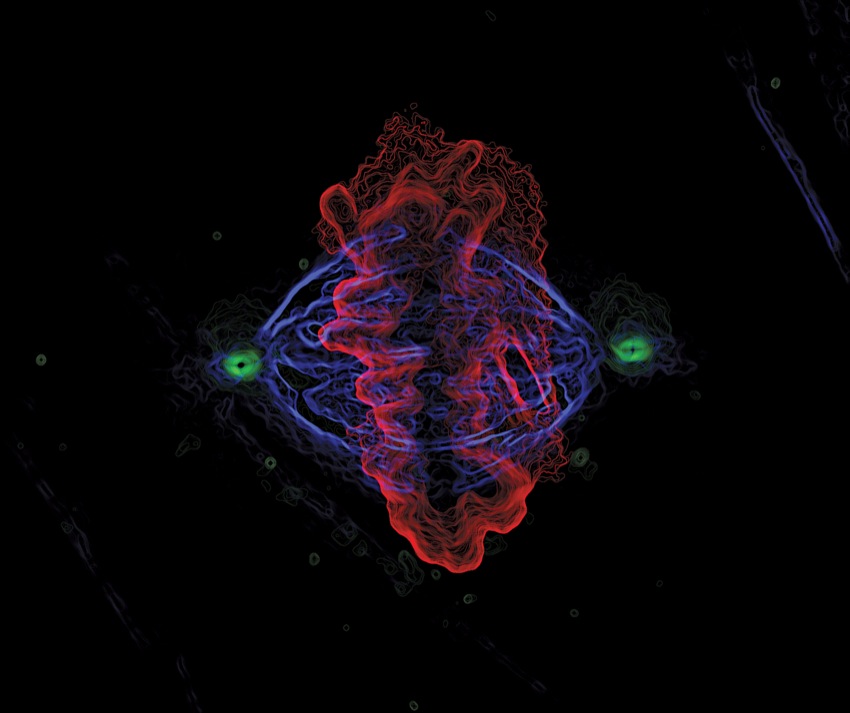
Focusing on microscopy
Fifteen years after Nordberg graduated from Wheaton, the lesson he learned from the setbacks—that science is unpredictable—keeps him grounded, focused and inspired in his career in microscopy, a technical field using microscopes to view objects at a cellular level.
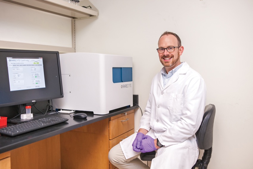
Nordberg, who has a Ph.D. in biomedical sciences from University of Massachusetts Medical School (UMMS), works as a senior technology development scientist at Seattle-based RareCyte.
RareCyte is a small, independent biomedical technology company that was spun off of the life-sciences microscopy company Applied Precision, when Applied Precision was purchased by GE Healthcare in 2011. RareCyte is dedicated to building technology that enables the analysis of rare circulating cells across the spectrum of disease, which can help answer difficult clinical questions involving cancer, prenatal diagnostics and infectious disease.
At the startup since 2011, Nordberg plays a key role in developing diagnostic tests for cancer and Lyme disease, and his work has led to two patents.
He first learned about Applied Precision while working as a course coordinator for “Analytical and Quantitative Light Microscopy” at the Marine Biological Laboratory in Woods Hole, Mass., from 2001 to 2008. The course, held every spring for 10 days, taught researchers advanced concepts in how to properly use their microscopes as an accurate measurement tool rather than just to see things bigger.
Companies sent their best equipment and application scientists to help teach the course, Nordberg said. “I got to know Paul Goodwin, a senior employee at Applied Precision. I wanted to work in research and development in the microscopy field, so I let him know of my intentions for when I graduated from UMMS.”
But Applied Precision CEO Ron Seubert had a different idea. He wanted Nordberg to work at his new company, RareCyte.
“When Ron explained that they were developing the technology for a new cancer diagnostic device, I jumped at the opportunity. I was brought in for an interview, and at the end I was asked—outright—whether I wanted to work at RareCyte or if I wanted to work at Applied Precision. The choice was mine,” Nordberg said.
As a technology development scientist at RareCyte, Nordberg bridges together the engineering, software and biology groups and works closely with Seubert.
“As a startup, we all wear many hats. It’s never boring,” he said. Some days he conducts biology experiments in the lab, while other days he finalizes the assembly and calibration of an instrument, or calls customers and coordinates a sale.
Describing Nordberg as methodical, detailed and creative, Seubert praised his contributions at RareCyte. “I hired Josh to be our voice, the one who could talk to customers and understand what they are trying to achieve,” Seubert said. “He’s done all of that—and more.”
Seubert says in addition to being a great communicator and listener, Nordberg has the scientific and technical know-how and diligence to ensure RareCyte’s novel technology is implemented properly at client sites. Plus, he is great to work with.
“You want to work with the smartest people who are nice—so you can get things done,” he said. “Josh is both of those.”
Cancer research
Nordberg has contributed to two patents during his time at RareCyte.
The first was for a project the company decided not to pursue. The second patent (number 9,039,999) is a source of great pride for the young scientist. Issued on May 26, 2015, the patent is for a core piece of technology that has implications for diagnosing and treating cancer, among other diseases.
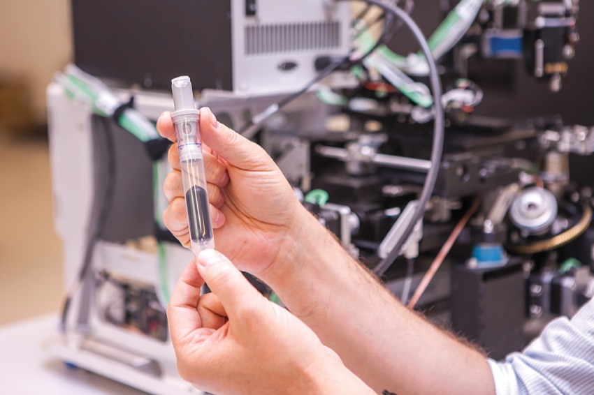
Nordberg’s team developed a kit that works in conjunction with RareCyte’s custom microscope and isolates all nucleated cells from a patient’s blood sample and puts them on slides. While the main focus of the company is the analysis of circulating tumor cells as a means for directing cancer therapy, RareCyte is also using the technology in the analysis of fetal cells and infectious bacteria.
The use of circulating tumor cells in the treatment of cancer differs from standard methods of diagnosis, which may involve a tissue biopsy to identify abnormal cells. “Biopsies can be painful and they carry risks such as infection,” said Nordberg, adding that some tissues are impossible to biopsy because of their locations. Another standard method of diagnosis is MRI imaging, but that process is expensive and time-consuming.
RareCyte’s method of a simple blood draw is more representative of the current state of the tumor, because old cells don’t last very long in the blood, he said.
“Circulating tumor cells offers a more real-time window into the progress of the cancer in a much lower-impact way for the patient.”
Blood collections can be performed weekly in most patients to help monitor treatment. Researchers can count the cancer cells; a lower number of cells indicates treatment success and a rising number indicates ineffective treatment.
“Our technology enables us to do what nobody else can. I worked on designing and fabricating one of the early prototypes and running the pilot experiments,” he said. “It was amazing to watch this product develop from concept, to a handful of prototype parts, to an injection-molded plastic part made by the thousands and used every day by our lab and now by other top-tier oncology researchers.”
His team also developed a device that works with their microscope to retrieve single cells for molecular and genetic characterization. By being able to isolate a single cancer cell, or a group of single cancer cells, the researcher can look for mutations in specific genes, or expressions of certain proteins on the surface of the cells, that may indicate how a patient could respond to a certain therapy.
“The hope is to avoid giving a patient a therapy that they will not respond to that could cause them to suffer unnecessary side effects. Also, the physician or researcher could use this to monitor changes in the tumor that could lead to a patient becoming resistant to a therapy—and help guide a change in therapy before any disease could recur,” he said.
Working to improve cancer diagnosis and treatment helps satisfy the part of Nordberg that once wanted to go to medical school. “It’s a great feeling to have every day, knowing that we developed this tool that can make a big impact on someone’s life and well-being.”
Targeting Lyme disease
The same method RareCyte researchers use to target cancer is now being applied to find rare bacteria, which include the bacterium that causes Lyme disease.
“Lyme disease is very difficult to diagnose. Symptoms can be so vague that it’s often missed in early stages when it is most easily treated,” said Nordberg.
Current diagnostics rely on detecting the patient’s immune response to the infection rather than directly detecting the organism causing the disease, he said. “The biggest problem with that approach is that it takes time for the immune system to react. The current tests are notoriously unreliable.”
When doctors can’t obtain a definitive result from the test, they are reluctant to treat, he said. “The side effects and consequences of long-term antibiotic use are well documented. What you are left with is a population of people who feel they aren’t getting the treatment they need.”
RareCyte’s blood test aims to be more sensitive and provide an early and more accurate and definitive diagnosis, he said.
In August 2014, the National Institute of Allergy and Infectious Diseases Small Business Innovation Research Program awarded RareCyte (in collaboration with the Western Connecticut Health Network Biomedical Research Institute) a two-year $600,000 grant to support the efforts to improve Lyme disease testing.
Enduring impact
Students often remember that one professor who helped shape their life’s work. In the case of Nordberg, it was the professor who found inspiration in a student.
“Josh was one of the reasons I knew I was at the right place,” said Morris, who joined the Wheaton faculty in 1998 and served as Nordberg’s advisor. “I was lucky to get a creative and thoughtful, intelligent and delightfully friendly young scientist in my laboratory as one of my earliest collaborators.”
For his senior thesis on embryonic ciliary growth kinetics, Nordberg utilized a newly acquired research-grade microscope. His time at Wheaton predated the digital imaging era (and Wheaton’s Imaging Center for Undergraduate Collaboration, or ICUC); he worked with then state-of-the-art equipment, including videotape and black-and-white video monitors to study cellular activity.
Nordberg’s experiments involved generating embryos and observing embryonic growth. He studied cellular appendages called cilia, which cells use like oars to move fluid and as antennae to listen for signals.
“He figured out how to measure cellular behaviors quantitatively and rigorously with no other students to show him how,” said Morris. “He observed behaviors that no one had observed before and that have profound implications for human disease.”
Specifically, Nordberg found that cilia grow in stages and don’t grow uniformly from beginning to end.
“He’d measure the lengths of the cilia as they grew and determined they grow in steps. They retract as the cells divide. They all grow to this pause, and continue to grow from there,” said Morris. “It’s very nice work.”
Nordberg innovated with his equipment to make these discoveries. He taped transparency screens to computer screens and figured out how to measure the length of cilia with time-lapse video. He’d take photographs of a TV tube in order to make the figures for his thesis.
His work on campus had an enduring legacy. The data he generated on sea urchin development helped launch other students on their research paths. His work and that of other students also provided the preliminary data that convinced the National Science Foundation to support creation of the ICUC. Some of this work is still in process today.
Nordberg presented his research at the American Society for Cell Biology’s annual conference in Washington, D.C., in December 1999. There, Nordberg met Greenfield “Kip” Sluder, a professor at UMass Worcester. Morris recommended Nordberg for a technician opening in Sluder’s lab. By that time, Nordberg had switched his interest in becoming a physician to wanting to pursue a career in research.
His work in Sluder’s lab went so well that Sluder ended up recruiting more Wheaton students, and Nordberg himself ended up circling back to the lab as part of his graduate studies.
Reflections on Wheaton
Nordberg credits Wheaton with preparing him for professional success.
His experience with Professor Morris showed him how much fun scientific research could be; and that the results of basic science research could eventually be applied to advances in medicine, he said. “I liked that connection.”
As a student, he worked hard, noted Professor of Chemistry Elita Pastra-Landis, who recalls him as a top-scoring student on her “Chemical Principles” exams and enthusiastic during early morning MCAT weekend study sessions.
Nordberg said he benefited from hands-on research and took multidisciplinary courses that broadened his lens when approaching scientific problems. “Wheaton helped round out my entire educational experience, in terms of learning how to learn rather than getting hyper-focused on one subject too early,” he said.
Wheaton also gave him confidence to work outside of his comfort zone. For example, he opted to take an advanced genetics course in graduate school when he could have coasted through a basic microscopy course. Also, his decision to work at RareCyte signaled a willingness to try something different.
“I don’t know that I would’ve had that attitude had I not had to take, say, an anthropology or religion class at Wheaton,” he said.